
Un dispositivo basado en un chip de bajo coste que se acopla a un smartphone está siendo desarrollado en la Harvard Medical School por investigadores del Massachussetts General Hospital para una detección rápida y precisa del cáncer y de otras enfermedades.
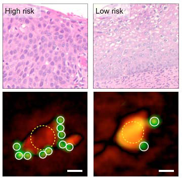
By quantifying the number of tumor-marker-targeting microbeads (green) bound to cells (lower images), the D3 system categorizes high- and low-risk cervical biopsy samples as accurately as traditional pathology (upper images). (credit: Massachusetts General Hospital Center for Systems Biology)
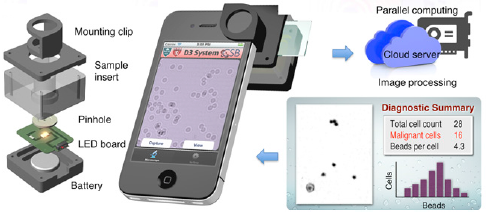
The D3 (digital diffraction diagnosis) system features an imaging module with a battery-powered LED light clipped onto a standard smartphone. It records high-resolution imaging data with its camera. With a much larger field of view than traditional microscopy, the D3 system is capable of recording data on more than 100,000 cells from a blood or tissue sample in a single image. The data can then be transmitted for analysis to a remote graphic-processing server via a secure, encrypted cloud service. The results can be rapidly returned to the point of care for viewing. (credit: Hyungsoon Im et al./PNAS)
No hay comentarios:
Publicar un comentario